BIOENGR 167L - Bioengineering Laboratory
BE 167L - Bioengineering Laboratory
Lab 7: Growth Kinetics and Protein Content (BCA Assay)
Prelab reading
Read the posted article from Current Protocols in Protein Science (2007) by Olsen and Markwell. (Read the following pages only: pgs. 1, 13-17. If you are curious about other methods of protein quantification you may read the entire article.) Please also read the instructions for the BCA assay kit you will be using to familiarize yourself with the reagents. A separate protocol adapted from these instructions is below for you to follow.
Peptide bonds (which exist between each amino acid in all proteins) reduce Cu+2 ions to Cu+1. The assay’s working reagent looks green in the beginning because of the copper. If you heat the protein/copper solution with the bicinchoninic acid (BCA), the peptide bonds also help to form a complex between two BCA molecules and the Cu+1 ion. This large molecule looks purple and absorbs maximally at 562 nm. There are a few specific amino acids (cysteine, cystine, tryptophan, and tyrosine) that also help form this purple complex, but if you heat the solution, the peptide bonds will do most of the work so that your signal will not depend heavily on the actual types of proteins present. It will rather be a fairly linear representation of total protein content. So taking a step back, what is the physical property that you should measure?
You will quantify the growth kinetics of your HeLa cells by two methods, cell counting on a hemocytometer and protein concentration. The protein content of different types of cells varies due to inherent size and activity. By using both of these methods on the same population of cells, you will be able to quantify the average amount of protein collected from a single 3T3 cell and use this general information to quantify cell concentrations in future experiments where cell counting is not an option.
You will use a RIPA buffer to lyse (break open) the cells and extract the proteins from the cells. (RIPA buffer derives its name from the original application for which it was developed: the radioimmunoprecipitation assay. While this isotopic assay method is rarely performed in laboratories today, the acronym for this lysis buffer formulation has endured in common use.) A combination of detergents breaks apart the cell membrane and cytoplasmic components and suspends them in the solution. The manual for using RIPA buffer can be found here. Once the cells are lysed and the proteins are released, enzymes may break them down from their natural or native state. You are not concerned about this break-down for this lab, but in the future if you wish to collect specific proteins from cells and plan to detect them using antibodies or other methods that rely on the whole protein being intact, you may add a Protease Inhibitor Cocktail to prevent proteolysis and maintain the protein’s native state or phosphorylation.
Once your proteins have been extracted, you will use the BCA assay to detect total protein concentration, that is the concentration of all types of proteins in the cells. On average, your population of cells will be uniformly sized and contain roughly the same amount of “stuff” or protein. You can use this measurement to represent how many cells you have, and over time you can track the proliferation of your cell population by measuring the protein content without actually counting the cells. Can you think of situations where counting cells might not be feasible?
For example, if you are growing your cells in a different environment, such as a porous scaffold or a gel, or even collecting cells from a whole tissue rather than culturing individual cells, you may not be able to collect whole cells for counting in a hemocytometer without breaking them apart into debris or losing some of them and decreasing your accuracy. You may also have too many samples and replicates to realistically count by hand. Using an assay like this can increase your accuracy of quantifying content and increase your throughput, particularly if you have a microplate reader (which we do!). This assay does not let you look at individual cells, locations of cells and how they are distributed within the culturing environment, or condition of the cells (healthy or sick, spread or rounded, dividing or quiescent). The BCA assay is only a measure of total protein. In later labs you will learn other assays that give you different types of information about your cells.
Watch the lab primer video.
Growth kinetics: counting and protein assay
Preparation
Reagents
- Complete medium from previous lab
- Sterile DPBS from previous lab
- Trypsin
- Your T25 flask and 12-well plate of cells from the previous lab
- RIPA buffer
- BSA protein standards
- BCA assay Working Reagent (WR)
Supplies
- Pipettes and tips
- Pipet-aid and serological pipettes
- 15 mL conical tubes
- 96 well plate to be used by the entire section
Equipment
- Phase contrast microscope
- Centrifuge
- Hemocytometer and cell counter
- Shaker or beaker with ice or ice water
- Microcentrifuge
- Plate Reader
Safety
Wear your lab coat, and gloves. Continue to practice good sterile cell culture techniques and proper biohazard waste disposal. Remember that RIPA buffer is a combination of detergents that lyses cells, and your body is composed of cells, so don’t come into prolonged contact with it, and wash it off right away if you do. The BCA assay working reagent is a very basic (pH) buffer, try not to contact it directly and wash off quickly if you do.
Procedure
You and your lab partner may start working on different parts of the procedure at once. One may begin the cell counting and protein extraction procedure, while the other begins preparing the standards and working reagent for the BCA assay. Read both sections below before you begin, and coordinate your responsibilities accordingly to complete this lab efficiently.
Cell counting and protein extraction
- Collect your flask and your 12-well plate and check them under the microscope. Estimate the confluency and cell density based on your experiments last week.
- Aspirate medium, trypsinize cells and collect cells from the well plate using the same protocol as last week. For specific volumes in a 12 well plate use the volumes listed for a 6 well plate except divide those by 2 (1 mL becomes 0.5 mL).
- Combine two wells into 1 tube so that you have a total of 6 tubes. You should have two tubes per condition.
- Centrifuge your tubes at 150 x g (1.5 rcf) for 5 minutes, aspirate the medium and resuspend your cells with 1 mL of DPBS for each of the tubes. As usual, do not start the centrifuge until the TA has checked to make sure that it is properly balanced.
- Take one tube from each seeding condition and count them on the hemocytometer. You may need to further dilute this tube prior to counting in order to count a reasonable amount of cells. Because you combined two wells together you will have to divide by 2 in order to get an average number of cells per well.
- Centrifuge your other tubes again at 150 x g (1.5 rcf on the other centrifuge) for 5 minutes. This rinses your cells of proteins in the medium and trypsin before you extract the protein from your cells.
- Aspirate the DPBS and add 200 μL RIPA buffer to the cell pellet to lyse the cells. Do not take more than needed. Pipette up and down to mix. Transfer this suspension to a properly labeled microcentrifuge tube.
- Put each of your microcentrifuge tubes (you should have 3) on ice or in ice water and shake gently for 15 minutes, then centrifuge the tubes in the microcentrifuge at 14,000 x g for 15 minutes (again check with the TA to make sure it is properly balanced). Note: During this time, you or your lab partner may trypsinize your T25 flask and passage those cells to a new 96-well plate (to be shared with other groups) with densities of 4,000 and 10,000 cells/cm2. You will have 4 wells of each cell density. Label them appropriately on the 96 well plate sheet your TA will have so you know which wells are yours. Seed a new T25 flask for next week.
- Without disturbing the pellet on the bottom of the microcentrifuge tubes (which may be very faint), carefully transfer the supernatant to a new, properly labeled microcentrifuge tube.
BCA assay
You will prepare your working reagent from reagent A and reagent B from the Pierce BCA kit. Although your TA will prepare the BSA standard stock solutions and dispense them to you, the protocol for preparing the standards is presented below for completeness.
A glass ampule of BSA at 2 mg/mL concentration is provided in the kit to make serial dilutions for a standard curve. Your TA used the following table to create the standards.
Table 1. Preparation of Diluted Albumin (BSA) Standards. Dilution Scheme for Standard Test Tube Protocol and Microplate Procedure. (Working Range = 20–2,000 μg/ml)
| Vial | Diluent Volume | Volume and Source of BSA | Final BSA Concentration |
|---|---|---|---|
| A | 0 | 300 μl of Stock | 2,000 μg/ml |
| B | 125 μl | 375 μl of Stock | 1,500 μg/ml |
| C | 325 μl | 325 μl of Stock | 1,000 μg/ml |
| D | 175 μl | 175 μl of vial B dilution | 750 μg/ml |
| E | 325 μl | 325 μl of vial C dilution | 500 μg/ml |
| F | 325 μl | 325 μl of vial E dilution | 250 μg/ml |
| G | 325 μl | 325 μl of vial F dilution | 125 μg/ml |
| H | 400 μl | 100 μl of vial G dilution | 25 μg/ml |
| I | 400 μl | 0 | 0 μg/ml (blank) |
- You have 9 standards and 3 unknowns. Create enough WR for you to have 200 µL of WR per sample. Prepare the WR by mixing 50 parts of BCA reagent A with 1 part BCA reagent B. Note: when mixing, the solution turns clear green but may go through a slightly cloudy phase at the very beginning. WR is stable for several days when stored in a closed container at room temperature. WR does not need to be protected from light during storage.
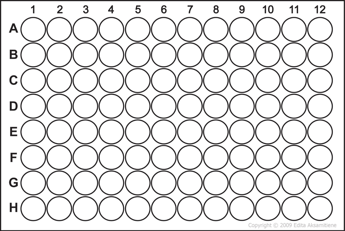
- Label your wells to be used on the 96 well plate. Your TA may ask you to share well plates.
- Dispense 25 µL of each standard from your TA into the appropriate well. Do not take more than is necessary.
- Dispense 25 µL of each of your unknown protein extract samples into the appropriate well.
- Add 200 µL of WR to each well and mix on the shaker.
- Cover and incubate at 37℃ for 30 minutes. Cool the well plate to room temperature
- Record the absorbance of each well and record the data related to your wells.
- At this point please also get data from two other groups, when you report your data, you will use your group plus two other groups data for statistical analysis. Don’t forget during your analysis that your protein lysate is from two wells!
